Copyright © 2022 Foshan MBRT Nanofiberlabs Technology Co., Ltd All rights reserved.Site Map
1. Research value
Corneal blindness is the second leading cause of blindness worldwide. At present, corneal allograft, artificial cornea and amniotic membrane transplantation are the most widely used methods for the treatment of corneal diseases. However, compared with the original cornea, these treatment methods have certain problems or defects. Therefore, the development of a therapeutic corneal scaffold using bioengineering and electrospinning technology to induce corneal stroma regeneration can effectively avoid the defects of the above clinical treatment methods, and has a very high research value.
2. Research content
①A novel structure controllable nanofiber collection device was designed and developed to realize the preparation of highly directional 3D PLGA nanofiber scaffolds, breaking through the limitation of traditional electrospinning technology which can only prepare 2D non-directional fibrous scaffolds. A composite corneal scaffold composed of collagen /PLGA nanofiber membrane/collagen was prepared by integrating electrospinning technology with compressed collagen.
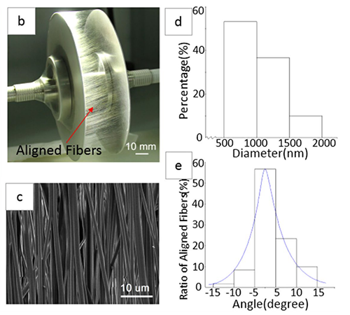
Figure 1.Electrospinning device and PLGA nanofibrous scaffold: (a) the main component of the device; (b) aligned fibers deposited between two parallel, thin plates; (c) an SEM image of a PLGA scaffold; (d) a histogram of fiber diameter; and (e) a histogram of fiber alignment.
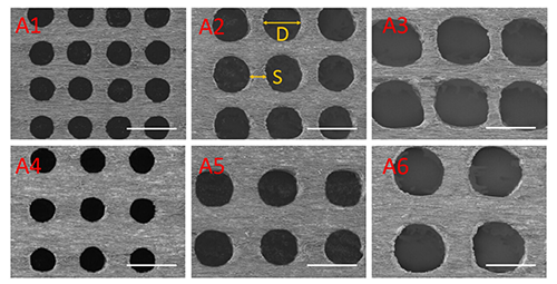
Figure 2. SEM images of laser-perforated, aligned PLGA scaffolds with different hole sizes (D) and spacings (S); A1-A6 are the samples shown in Table 1. The scale bar is 200μm.
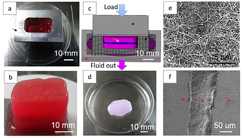
Figure 3. The hybrid scaffold production process
②The corneal stromal cells were encapsulated in collagen and the corneal epithelial cells were inoculated on the surface of the composite scaffold to achieve the co-culture of the two kinds of cells in vitro.
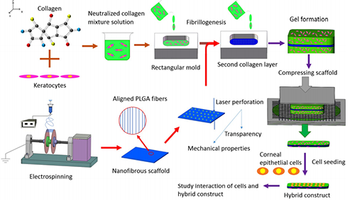
Figure 4. The schematic illustration of the experimental process.
③The PECL microfiber scaffolds were fabricated by near field electrospinning technique and simulated the natural extracellular matrix structure of cornea. It is reported for the first time that corneal stromal cell phenotype and induced regeneration of corneal stromal cells can be maintained under the synergistic action of orthogonal-oriented topological structure and specific chemical factors.
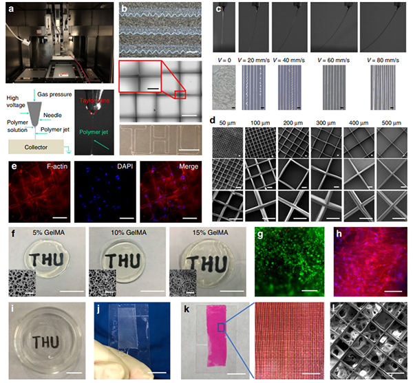
Figure 5. Fabrication of characterization of the fiber hydroge
Fabrication of characterization of the fiber hydrogel. a The custom-made direct writing device, a schematic representation of direct writing, and a polymer jet being ejected from a Taylor cone. b Images of the obtained curly lines (Scale bar is 500 μm), grids (Scale bar is 500 μm and the enlarged imageis 150 μm), straight lines, and a specifically designed pattern (Scale bar is 10 mm). c Images of stable jet deposition at different the moving speeds of thesubstrate in the specific conditions. The scale bar is 100 μm. d SEM images of the grid scaffolds with fiber spacing of 50–500 μm under different magnification. The scale bar is 50 μm. e LSSCs inoculated on the grid scaffolds can be induced to grow along the fiber direction, cells were stained withphalloidin (red) and DAPI (blue). The scale bar is 100 μm. f The macroscopic and microscopic morphology of the formed GelMA hydrogels with 5%, 10% and 15% concentrations. The scale bar for macroscopic images is 10 mm, for the SEM images is 80 μm. g The viability of LSSCs within the 5% GelMA hydrogel. Live cells are visualized with green, and dead cells appear red. The scale bar is 500 μm. h The cytoskeleton (red) and DAPI (blue) staining ofLSSCs within the 5% GelMA hydrogel. The scale bar is 100 μm. i–l The fabricated 100 μm spacing grid fiber-reinforced GelMA hydrogel. The scale bar in i–k is 10 mm, the enlarged image in k is 1 mm, l is 100 μm. The staining in e, g, and h were repeated three times independently with similar results
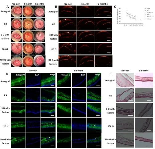
Figure 6. Fiber hydrogel engraftment and stromal matrix synthesis and regeneration in rat cornea in vivo
3. Relevant Academic Achievements’ Link
[1] Kong B , Chen Y , Liu R , et al. Fiber reinforced GelMA hydrogel to induce the regeneration of corneal stroma[J]. Nature Communications, 2020, 11(1).
Link:https://www.nature.com/articles/s41467-020-14887-9.pdf
[2] Kong B , Sun W , Chen G , et al. Tissue-engineered cornea constructed with compressed collagen and laser-perforated electrospun mat[J]. Scientific Reports, 2017, 7(1):970.
Link:https://www.nature.com/articles/s41598-017-01072-0.pdf
[3] Kong B , Mi S . Electrospun Scaffolds for Corneal Tissue Engineering: A Review[J]. Materials, 2016, 9(8):614.